Prepare the plasma
≈60 mLblood/≈30 mLPRP + PPP
Collect the patient’s blood and separate the plasma using the selected preparation system. Both PRP and PPP are carried forward.
Tubes illustrate one preparation method.
THE EXOSMART™ SYSTEM
The patient’s own PRP and PPP, processed together through hollow-fibre ultrafiltration.
exosmart adds a concentration step after centrifugation. Water and permeable solutes leave through the membrane; retained platelet and plasma components remain together in a smaller volume.
Inside the system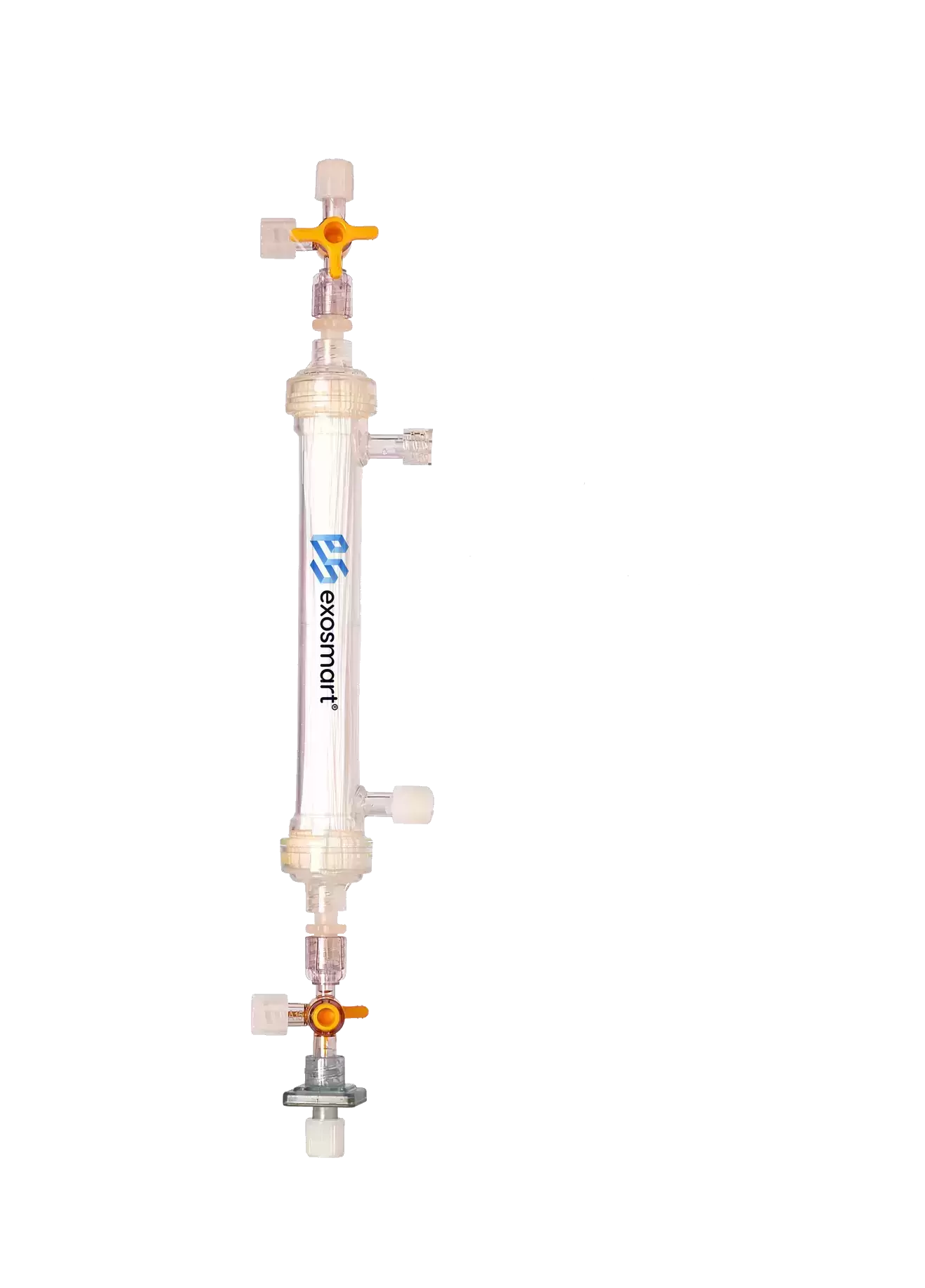
01 / THE STARTING MATERIAL
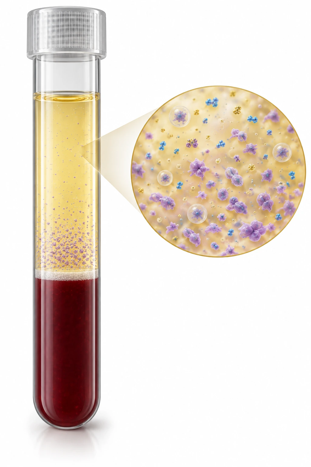
PRP supplies platelets and their associated signals. The surrounding plasma carries vesicles, soluble mediators and proteins involved in transport, regulation and matrix formation.
Supplies platelets and their stored and membrane-associated mediators.
Contributes circulating signals, carrier and regulatory proteins, and matrix-forming proteins.
We collect both fractions before concentration. Each preparation reflects the patient’s plasma and the centrifugation method used.
02 / FROM BLOOD TO CONCENTRATE
Collect the patient’s blood and separate the plasma using the selected preparation system. Both PRP and PPP are carried forward.
Tubes illustrate one preparation method.Plasma moves along the hollow fibres. Water and permeable small solutes cross the membrane while retained components concentrate.
Time for the concentration step.The retained preparation is collected in a syringe, ready for the next stage of the clinical workflow.
Final volume depends on the protocol.Typical preparation volumes are shown. The concentration step takes approximately 3–5 minutes after blood collection and centrifugation. Haematocrit and the preparation protocol influence the volumes and timing. Follow the current manufacturer’s Instructions for Use for device operation.
03 / INSIDE THE DEVICE
The syringes move plasma back and forth along the hollow fibres. This movement along the membrane surface is called tangential flow.
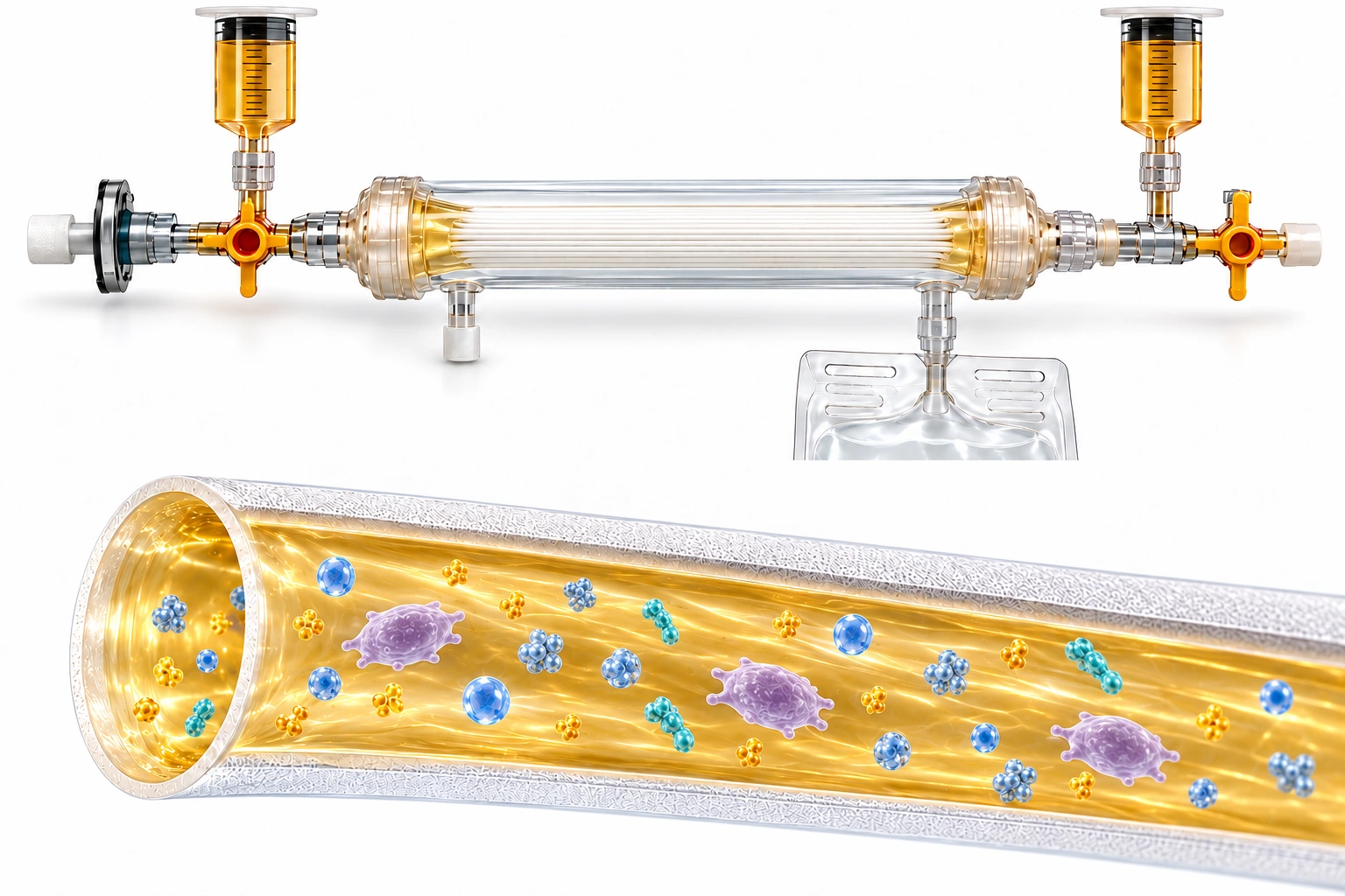
Inside the fibresPlasma moves back and forth along the membrane.
Across the membraneWater and permeable small solutes pass into the collection compartment.
In the preparationRetained components concentrate as fluid volume falls.
Platelets, vesicles and retained plasma proteins concentrate in the remaining fluid. Retention also depends on molecular form and binding to carrier proteins.
Water and permeable small solutes leave into the collection bag. The nominal cut-off characterises membrane permeability; molecular form also influences passage.
The final volume is approximately one fifth of the starting plasma volume. Laboratory testing measures the concentration and recovery of individual components.
04 / THE BIOLOGY WE KEEP TOGETHER
The Complete Autologous Plasma Secretome brings together vesicles, platelets, soluble signals, carrier and regulatory proteins, and matrix-forming proteins. Select each layer to see how it contributes to the biological environment.
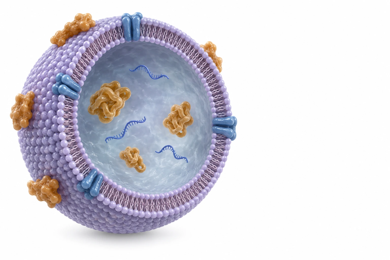
Extracellular vesicles carry proteins, lipids and RNA. Their surface and surrounding proteins also influence interactions with recipient cells.
exosmart concentrates vesicles together with platelets and the surrounding plasma components.
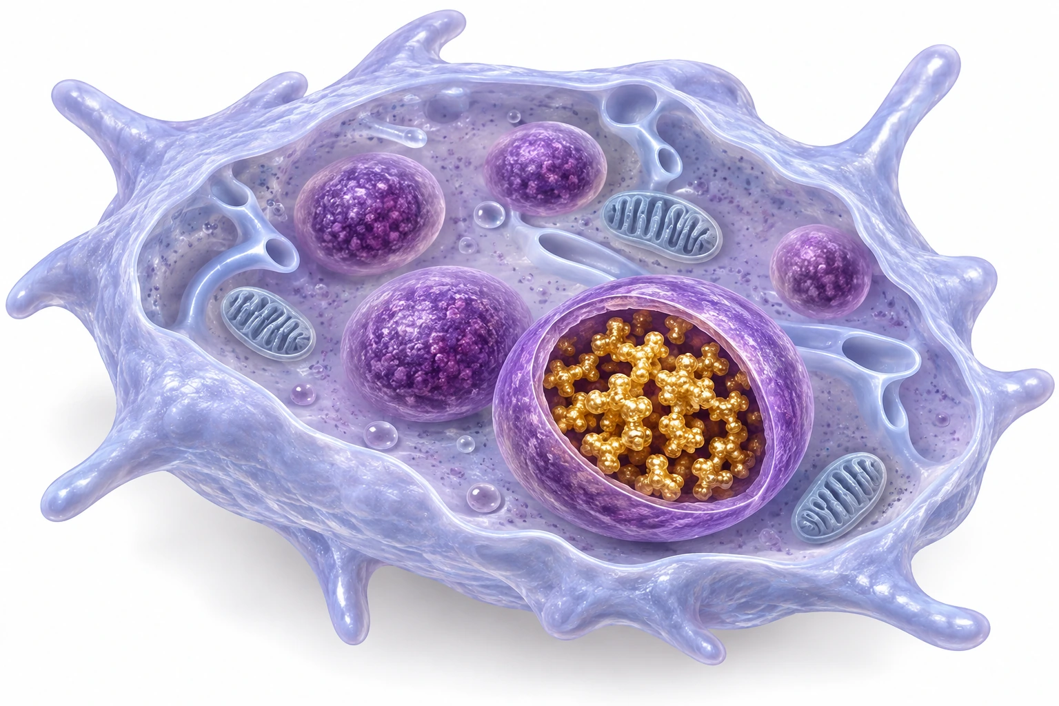
Platelets contain granules and membrane-associated proteins. They respond to their biological environment and contribute platelet-associated signals.
Platelet granules store mediators that can be released as platelets respond to local signals.
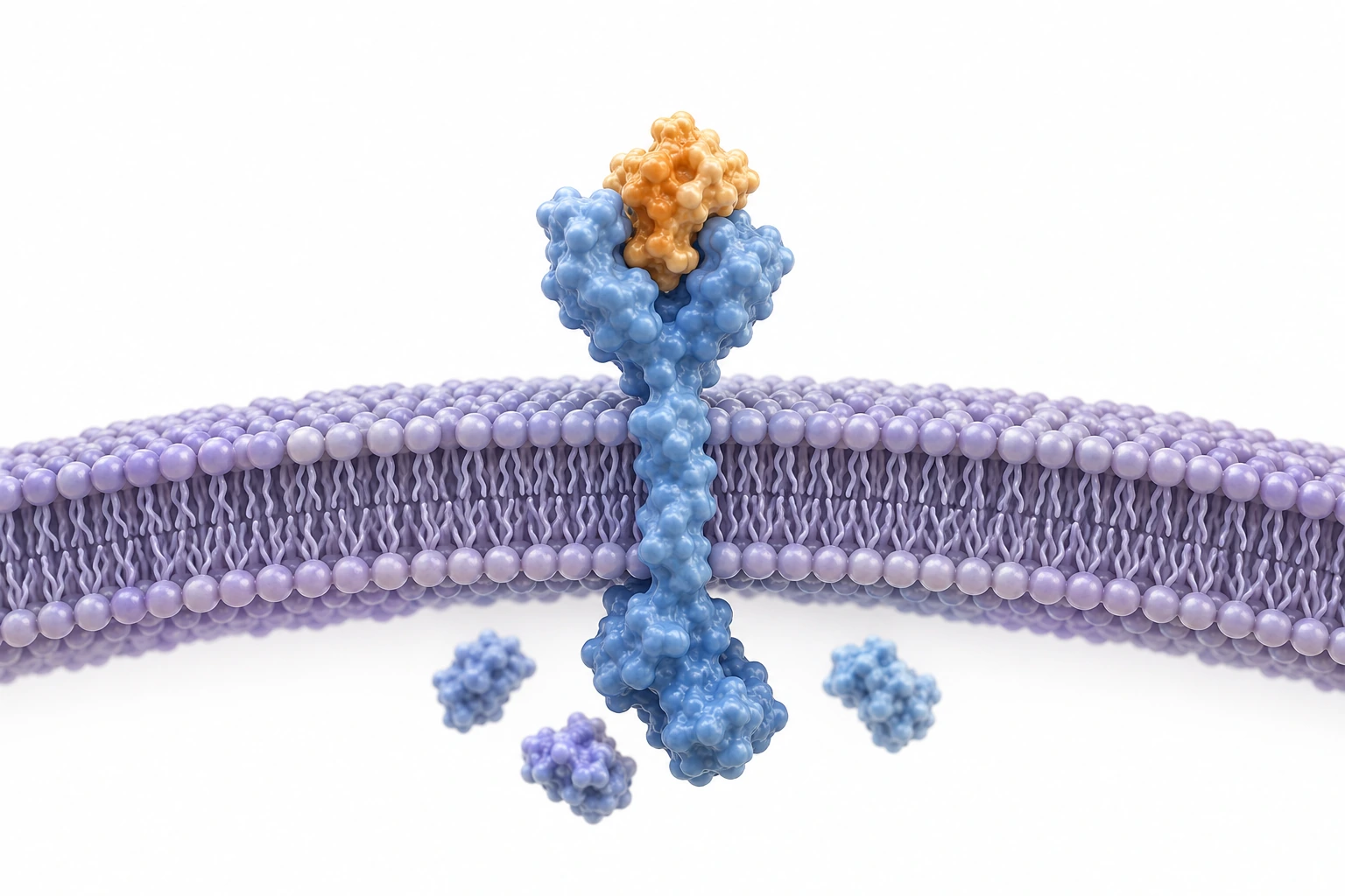
Growth factors and other soluble mediators interact with cells that carry the relevant receptors. Different signals have different biological roles.
PDGF-AA, PDGF-AB/BB, VEGF, TGF-β1 and EGF have been measured in the final preparation.
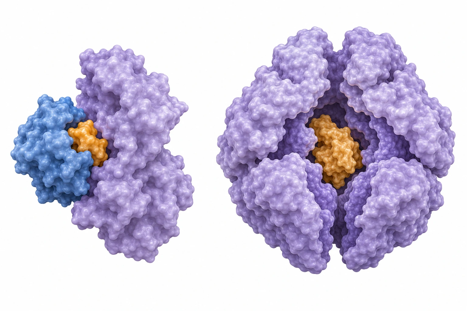
Plasma proteins transport and bind biological signals. Carrier complexes influence signal availability, while regulatory proteins help control biological activity.
The illustration shows carrier and regulatory protein concepts, including IGF-binding proteins and α2-macroglobulin.
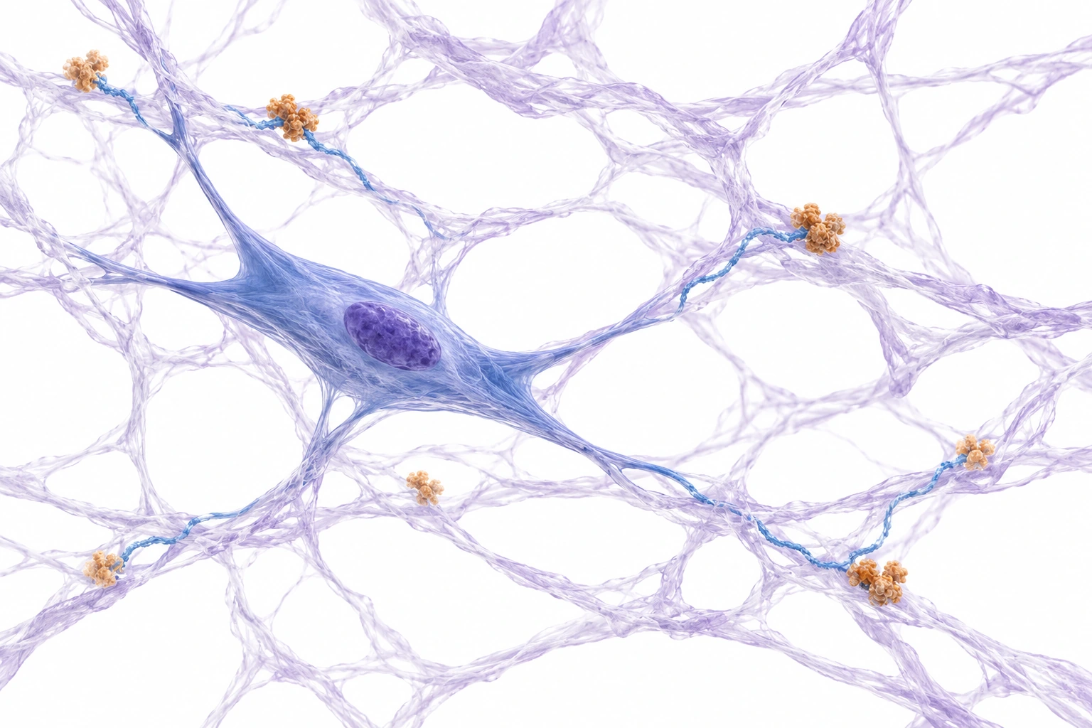
Fibrinogen and fibronectin participate in the local matrix. Fibrin forms when coagulation conditions trigger its assembly.
The illustration shows the matrix these proteins can form under appropriate local conditions.
These components interact within the same plasma preparation. The Science page connects their biological roles with laboratory findings and clinical observations.
05 / LABORATORY CHARACTERISATION
Particle tracking, microscopy, marker testing and growth-factor assays describe the preparation from complementary perspectives: size, structure and molecular composition.
Science & evidenceNanoparticle tracking analysis measures light-scattering particles. The laboratory reported 5–30 × 10¹² particles/mL across four finished preparations, with median diameters of 127–178 nm.
NTA counts light-scattering particles across the sample, including vesicles and other plasma particles such as lipoproteins.
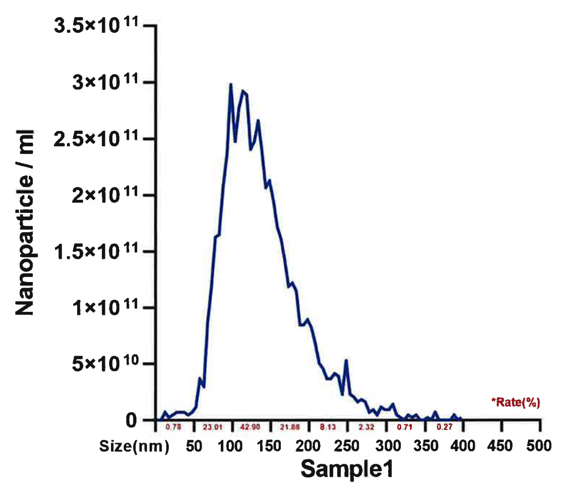
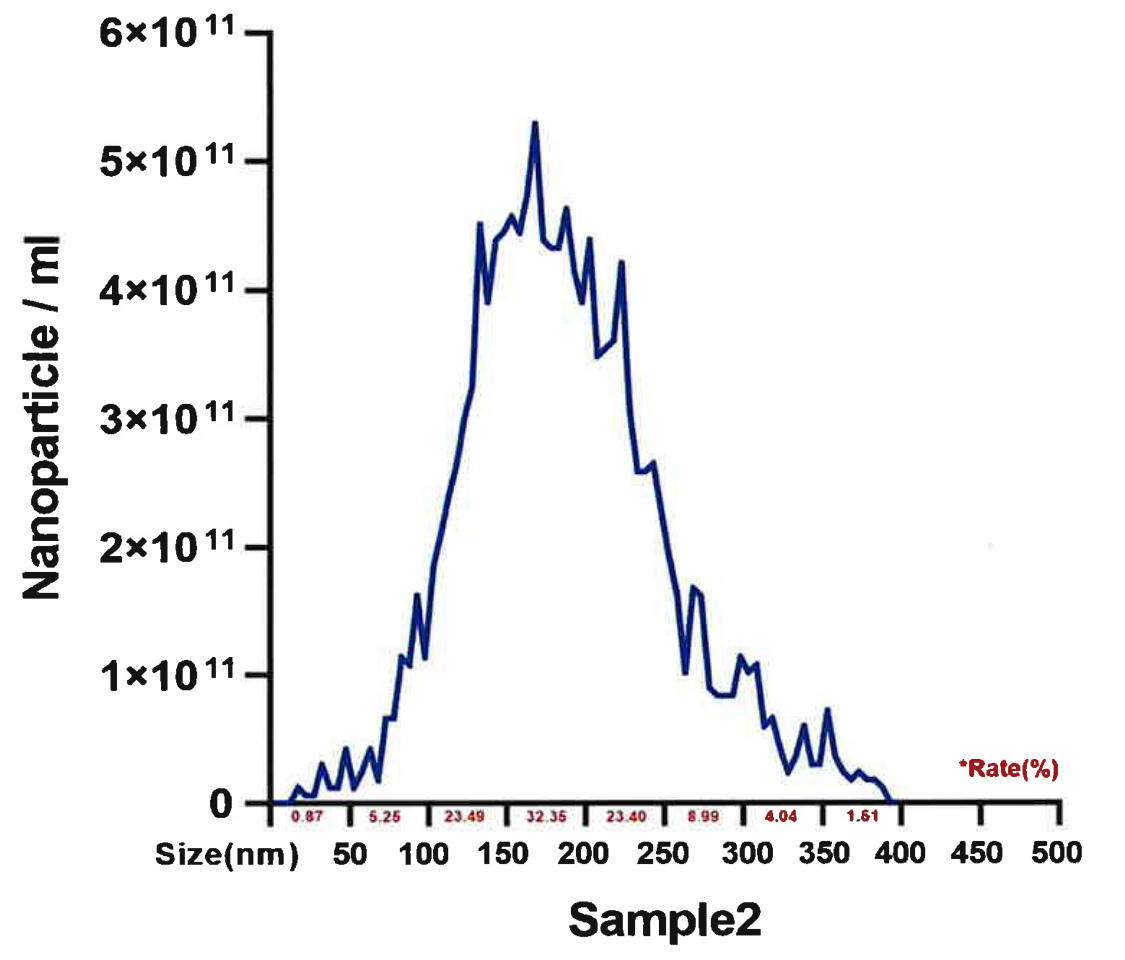
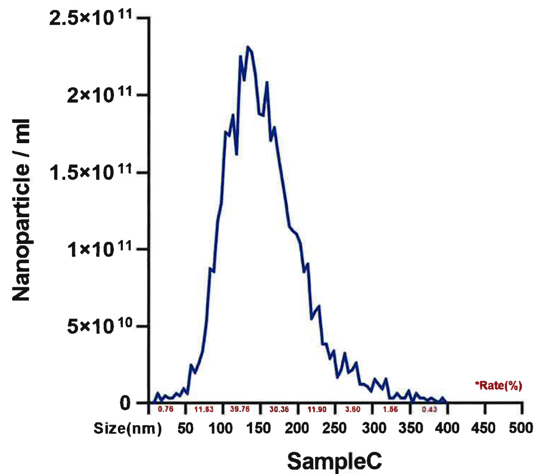
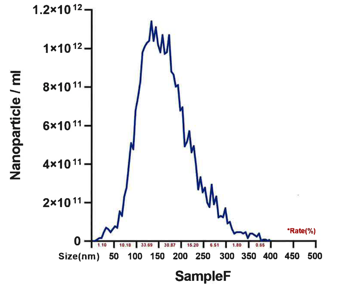
Source: StemExoOne, four finished preparations; original curves reproduced from Scientific Foundation, slide 32. Each graph retains its original vertical scale. Headline concentrations are laboratory-reported totals.
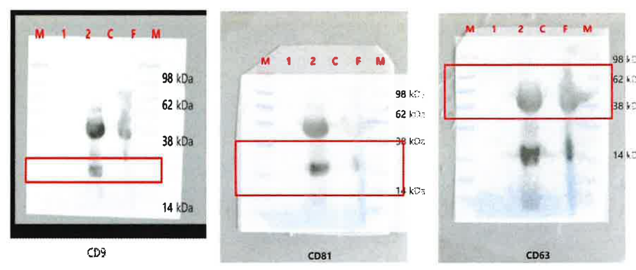
Western blot and flow cytometry identified the EV-associated markers CD9, CD63 and CD81 in the analysed preparations.
Read alongside microscopy and particle tracking, these findings add molecular detail to the characterisation of the preparation.
Three donor sets were assessed by a third-party laboratory using Luminex xMAP / Bio-Plex 200. The ranges show variation between donors.
| Growth factor | Observed range |
|---|---|
| PDGF-AA | 3,303–6,153 |
| PDGF-AB/BB | 1,428–3,936 |
| VEGF | 66.8–157.5 |
| TGF-β1 | 23,331–40,665* |
| EGF | 164–413 |
Source: Woongbee Testing Center, report 2025-027-L; three donors, triplicate wells. *The highest TGF-β1 value was extrapolated above the assay standard curve and is an estimate.
06 / PREPARATION IN PRACTICE
Follow the original exosmart preparation film, from the assembled circuit to collection of the concentrate.
07 / DOCUMENTATION & TRAINING
Ask our team for the current device documentation, analytical reports and clinical training for your specialty.
exosmart is a sterile, single-use hollow-fibre ultrafiltration device manufactured by Medica S.p.A.