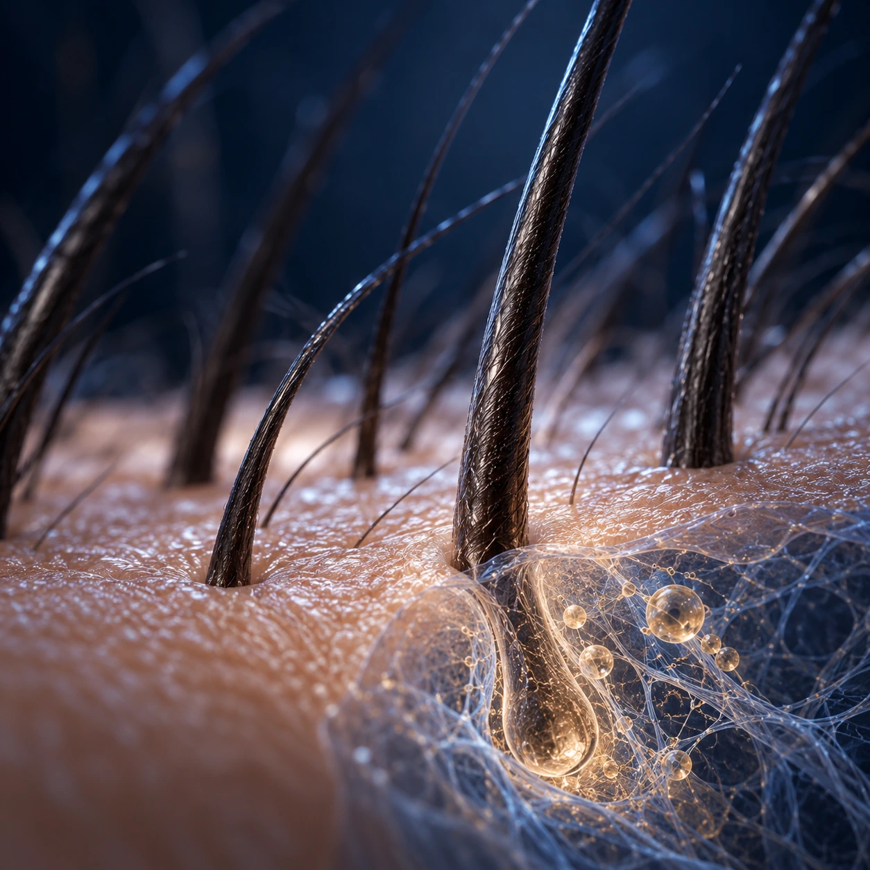
Follicle signalling
The dermal papilla and surrounding cells coordinate the hair cycle.

APPLICATIONS / HAIR & SCALP
Follicle biology, scalp assessment and documented follow-up for clinicians managing pattern hair loss.
THE TISSUE & THE PREPARATION
In pattern hair loss, assessment starts with diagnosis, follicular miniaturisation and scalp condition. The biological rationale considers dermal papilla signalling and the blood vessels and cells surrounding the follicle.
exosmart processes PRP and PPP together to carry platelet-associated signals, vesicles and plasma proteins into one preparation. The hair restoration handbook covers patient assessment, use alongside other therapies and follow-up timed to the hair cycle.
The dermal papilla and surrounding cells coordinate the hair cycle.
Vesicles, growth factors, carriers and regulatory proteins contribute different biological functions.
Repeat photography and trichoscopy keep the assessment tied to the same scalp area.
CLINICAL FOCUS
Androgenetic alopecia and female-pattern hair loss form the core clinical teaching.
Scalp assessment, current therapies and the patient’s history are considered together.
Specialist material addresses the scalp and perioperative tissue environment alongside the surgeon’s operative plan.
EXOSMART CASE SERIES · 10 PATIENTS · 90 DAYS
Dr V. Hernández Machado’s prospective case series followed 10 patients at baseline, day 30 and day 90. Of the nine patients who returned satisfaction forms, seven were very satisfied and two were satisfied. Photography and trichoscopy documented the follow-up; satisfaction was the reported outcome.
ASSESSMENT & FOLLOW-UP
The intervals below are educational follow-up guidance, not the schedule of a specific study. Record a baseline and adapt follow-up to the condition, procedure and patient. Refer to the specialty handbook for treatment planning.
Document the diagnosis, treatment area and photographic or trichoscopic baseline.
Review recovery, scalp condition and the wider management plan.
Make the first objective hair assessment using repeat images and trichoscopy.
Compare the findings with baseline and decide whether further treatment is appropriate.
What to record Standardised images · trichoscopy · scalp status · patient experience
PROFESSIONAL EDUCATION
EXOSMART ACADEMY · OCTOBER 2026
Biology, clinical evidence, assessment and follow-up for hair.
Written by Mihail Blagoev, co-founder of exosmart and Global Regenerative Group. Use the companion Evidence Compendium for study detail and the Pocket Card for a concise clinical reference.
Read the handbook All hair resourcesYOUR PRACTICE
Contact us for device preparation training, scientific documentation and education for your specialty.